

Silicone Plastinates
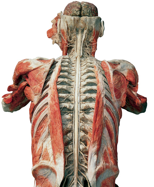
 Silicone plastinates are plastinated specimens from real human bodies. They include almost every body part — from organs to whole-body specimens. Their unique three-dimensional complexity illustrates the intricate structure of the musculoskeletal system in the inner organs as well as their relative position to each other.
Gunther von Hagens has shaped the whole-body plastinates so that they clearly show how the body is constructed and how it performs different tasks. Other whole-body plastinates have been dissected in ways that highlight different body systems, such as muscles, internal organs, or nerves and blood vessels.
Sheet (Slice) Plastinates
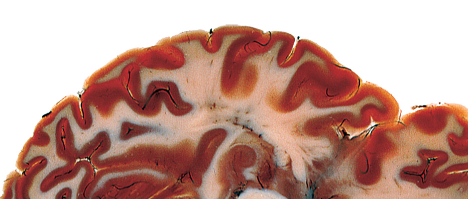
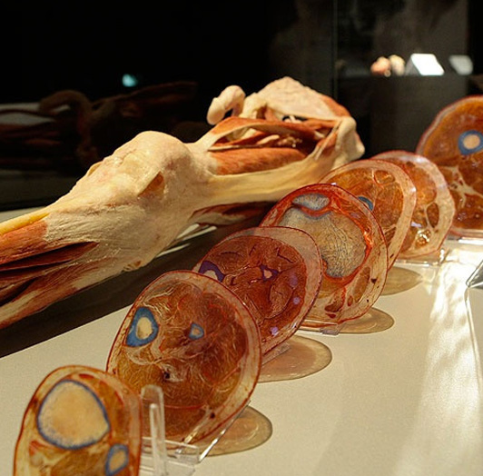
 Sheet (slice) plastinates are 1.5 mm thin slices of real bodies, impregnated with resins and subsequently hardened. In comparision to well-known diagnostic pictures of computer tomography (CT) and magnetic resonance tomography (MRT), sheet plastinates illustrate anatomical structures, in color, either transparent or translucent. The intricate world of macroscopy is revealed, as areas of interest can be studied down to the finest tissues, at the microscopic level. Being able to study the human body in the shape of perfect slices has been an anatomist’s dream for centuries as these slices illustrate the bodyss compact anatomy in an unaltered manner.
The method of plastination now, for the first time, facilitates the production and preservation of thin, transparent body slices of unparalleled transparency, vibrant colors, and size.
Blood Vessel Configuration
 Blood vessel configurations are perfect castings of the inner blood vessels, body parts and whole bodies. A revolutionary technological process, consisting of injection, curing and removal of remaining tissue, enables us to produce specimen that demonstrate precisely even the smallest and filigree blood vessels. They are created by injecting liquid colored plastics into the vessels. Once this plastic hardens, it assumes the shape of the vessels. Then, the surrounding tissue is removed, both mechanically and with the help of enzymes. A vessel configuration, therefore, solely consists of vessels that have been injected with plastic.
Blood vessel configurations are three-dimensional and illustrate the complex work of blood vessels in the human body or body parts.
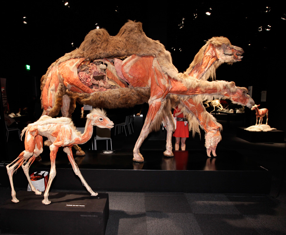
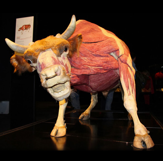
Animal Specimen
Animals have fascinated me all my life
 As a child, I was enthralled by the small animals I encountered in the woods.
The first specimens I dissected were beetles, frogs, and other small animal corpses that my friend, Dietrich and I found during our jaunts to the woods. These deaths which were so random and yet so normal must have colored my view of death and shaped my thoughts on mortality, preparing me psychologically for my career as an anatomist.
My childhood years were filled with a certain awe for nature and the varieties of life that populated it. But in my teenage years, my interest in biology was replaced by an interest in electronics and space. I became the resident expert on all things related to Sputnik, and soon in the gadgets I saw in early James Bond films.
Later as an adult, I renewed my relationship with animals by frequently visiting zoos and aquariums. The larger than life animals I admired—giraffes, elephants, and gorillas—were filled with a controlled grace that I found wondrous.
They lumbered, they sauntered, they ambled, their elegance so surprisingly disproportionate to their size. In the last decade, I have traveled to Africa and Antarctica to see up close the creatures that had captured my childhood imagination.
In an accelerated technological age, when our environments are fashioned from steel and concrete, being in close proximity to animals—both domestic and wild—return us to authenticity. Outside of the rainforests and flora, they and we are the last remaining pieces of nature. They are our co-habitants on this spinning blue globe. This exhibition, ANIMAL INSIDE OUT, is both a celebration and an homage to animals both familiar and rare.
Click here to learn more about animal plastination and the Animal Inside Out program.
Dr. Gunther von Hagens
Anatomist, Inventor of Plastination and
Creator of ANIMAL INSIDE OUT, a Body Worlds Production
February 2013
Gunther von Hagens found it necessary to establish the Institute for Plastination (IfP) in 1993 because the space and technical facilities available at the University of Heidelberg were no longer adequate for the growing demands of Plastination. The IfP in Heidelberg was where the techniques for preparing whole-body plastinates and transparent slices of whole bodies were perfected. The complexity and work involved in preparing these specimens far exceeds the capacity of most interested institutes. Preparing a technically correct, whole-body plastinate does, after all, require 1000 to 1500 man-hours.
The aim of the IfP is to produce human specimens and make them available both for basic and continuing medical training as well as for the general medical education of the public. The specimens are prepared solely for this purpose and only passed on directly to recognized educational and research establishments and scientific museums, but not to private individuals or dealers.
The objectives of the IfP can be summarized as follows:
- Improving overall anatomical instruction
The IfP produces high-quality educational specimens for anatomical instruction at universities and other teaching institutions. - Improving awareness of medical issues, particularly among the general public
The IfP produces plastinates aimed at educating non-medical professionals and restores public access to the anatomy of the human body. - Popularizing and developing plastination techniques
The IfP disseminates plastination expertise around the world, allowing other teaching institutions to profit from this unique process. The IfP also pursues scientific objectives and strives continually to develop and refine the techniques of Plastination and the resulting anatomical specimens. It is aided in these endeavors by visiting scientists and scholarship holders from national and international universities.
- Improving overall anatomical instruction
There are now more than 400 plastination laboratories in 40 countries around the world using Plastination to prepare specimens for academic study. Despite all of the progress made to date, the need for further research is immense. Tests need to be performed, for instance, on new polymers that could be used to retain the color of tissues and to improve plastination results for specimens such as the eye, which are difficult to preserve.
Every two years, participants at the International Plastination Conference have the opportunity of exhibiting the plastinates that they have produced. In addition, the “International Society for Plastination” and its publication “The Journal of the International Society for Plastination” provide additional forums for experts in the field to exchange information concerning advances in the scientific application of the process. Current issues include how slice Plastinates can be used to show complex systems such as the blood supply to the bones of the wrist or how to display subtle structures such as the muscles and nerves surrounding the prostate. These tissues are critical for proper sexual functioning and understanding them is an extremely important means of obtaining precision when planning delicate surgical procedures.
Sesneber International is a distributor for all Gubener products
The Anatomical Teaching Specimens are for purchase and use by only and strictly medical teaching institutions. All shipments are sent directly from Gubener Plastinate GmbH (Germany) to the purchasing medical institution. We welcome inquiries by medical distributors but please keep in mind that the customer’s full contact information must be declared at the time of the request for the price of the products.
If you are located anywhere in the Middle-East (with the exception of Iraq and Iran), please contact Sesneber International at the address listed below for price quotes and additional information:
Sesneber International
P. O. Box 87437
Riyadh 11642, Saudi Arabia
Phone: +966-11-454-1222
Fax: +966-11-453-7890
E-mail: info@sesnebe.com
Please Note: Sesneber International cannot supply any Anatomical Teaching Specimens from Gubener Plastinate GmbH to the Republic of IRAQ or IRAN. For these two specific regions, please contact Gubener Plastinate GmbH directly at: info @ plastination.com or visit their website at http://www.vonhagens-plastination.com/
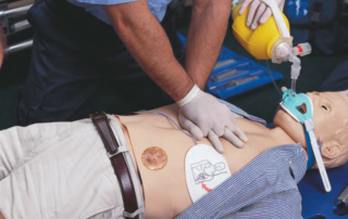
Medical Education and Simulation
Medical simulation and training models; real human and animal plastinated specimens; osteological models and specimens and educational media. Consultancy services and turnkey solutions for set up of medical simulation centers, including mobile facilities, emergency training centers and effective classroom design.
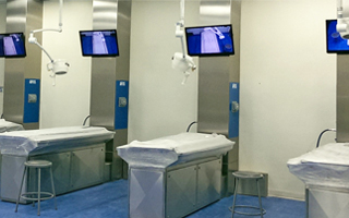
Anatomical Products & Services
Complete range of anatomical products, including anatomical models, real human and animal plastinated specimens, anatomy lab equipment and furniture. Consultancy services and turnkey solutions for set up of anatomy labs, including setup of audiovisual equipment.